fluid mosaic model animation mcgraw hill
DNA animations by wehitv for Science-Art exhibition 6 Steps of DNA Replication From DNA to protein - 3DLeading strand vs. The fluid mosaic model was proposed by SJ.
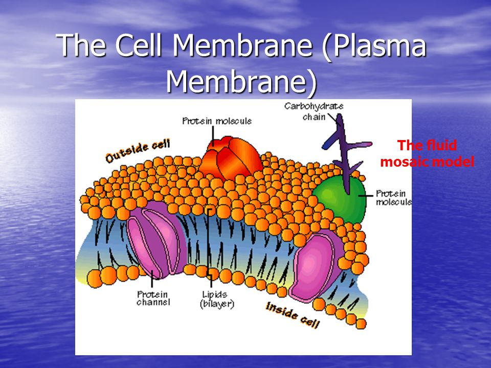
The Cell Membrane Plasma Membrane The Fluid Mosaic Model Ppt Download
Read Book Glencoe Mcgraw Hill Biology.

. Cell membrane overview and fluid mosaic model. This looks at the structure of cell membranes giving a snippet of the evidence supporting the Fluid Mosaic Model. Plasma membranes range from 5 -10 nm in thickness.
We would like to show you a description here but the site wont allow us. According to their model cell 3 membranes are composed of a lipid bilayer with globular proteins embedded in the bilayer. C H H H H H.
This is the currently selected item. Membranes also form specialized compartments within the cell. This model explains the structure of the plasma membrane of animal cells as a mosaic of components such as phospholipids proteins cholesterol and carbohydrates.
Describe the Fluid Mosaic Model of membrane 2 structure. Phospholipid bilayer hydrophilic heads point outward and hydrophobic tails point inward. Kristin has taught college Biology courses and has her doctorate in Biology.
To run the animations you must be in Slideshow View. Cholesterol molecules stabilize the membrane. The phospholipid bilayer gives fluidity and elasticity to the membrane.
The proportions of proteins lipids and carbohydrates in the. Objective 13 In 1972 Singer and Nicolson proposed the proposed the Fluid Mosaic ModelFluid Mosaic Model of of membrane structure. The fluid mosaic model explains various observations regarding the structure of functional cell membranesAccording to this biological model there is a lipid bilayer two molecules thick layer consisting primarily of amphipathic phospholipids in which protein molecules are embedded.
Cell membrane overview and fluid mosaic model. Such intracellular membranes help shape many of the morphologically distinguishable structures organelles for example mitochondria ER Golgi secretory granules lysosomes and the nucleus. Up to 24 cash back animations.
Lagging strand Chapter 7 Fluid Mosaic Model of the Cell Membrane Biology in Focus Chapter 1. A short video on the Fluid Mosaic Model of the cell membrane. You may see blank slides in the Normal or Slide Sorter views.
Membranes localize enzymes function as integral elements in excitation-response coupling and provide sites of. Introduction - Evolution and the Foundations of Biology Gel Electrophoresis. Attached peripheral and integral proteins serve as receptors channels and carriers.
Use the buttons on the animation to play pause and turn audiotext on or off. PowerPoint Presentation Last modified by. St Aidans Church of England High School Harrogate.
The Fluid Mosaic Model states that the cell membrane is. These components give a fluid character to the membranes. Animation Please note that due to differing operating systems some animations will not appear until the presentation is viewed in Presentation Mode Slide Show view.
Once you have used any of the animation functions such as Play or Pause you must first click on the slides background before you can advance to the next slide. The extracellular matrix and cell wall. 3 fatty acid molecules c c 0 c c I glycerol molecule I triglyceride molecule 3 ester bonds 3 water molecules 3 H20.
Caroline Mortimer Created Date. Each phospholipid has a hydrophilic head pointing. Singer and Garth L.
All animations will appear after viewing in Presentation Mode and playing each animation. The mosaic model of membrane structure describes the structure of the plasma membrane as a mosaic of components including phospholipids proteins carbohydrates cholesterol and proteins that gives the membrane a fluid character.